doi: 10.15389/agrobiology.2012.2.69eng
УДК [636.52/.58+636.028]:574.24:591.4
ELEMENT STATUS AND MORPHOFUNCTIONAL STATE OF REPRODUCTIVE ORGANS IN HENS AND MAMMALS UNDER THE INFLUENCE OF CADMIUM LOAD
S.A. Miroshnikov, S.V. Lebedev, A.I. Vishnyakov
On laboratorial rats and hens of the Highsex Brown the authors studied the influence of cadmium toxic doses on metabolism of chemical elements (Mg, Са, Se, Zn, Ag, I, K, P, Sr, As, Al, Pb) and morphofunctional state of reproductive organs. The addition of cadmium to ration leads to the disturbance in metabolism of chemical elements both in whole organism and in reproductive system, that accompanied by the reduction of selenium content and the decrease of functional activity of reproduction organs
Keywords: rats, hens, mineral exchange, cadmium, toxicity, reproductive system.
Environment protection from heavy metal pollution is one of the most pressing environmental problems. Elevation of toxic waste causes a damage to human health at direct contact or through contaminated food and water. Migration of toxic elements results in their bioaccumulation in water, soil, feed, animals and human (1). Ecological disturbance provokes the raise of morbidity and murrain in farm animals and wild species along with the reduce in their productivity. A prolonged exposure to toxic substances causes pathological changes in the body – metabolic disorders, suppressed immunity, neurohumoral dysfunction, and abnormal structure of organs and tissues, etc. (2, 3).
Cadmium is one of the most toxic heavy metals considered as a highly dangerous substance (2-hazard class) according to domestic sanitary rules and regulations (SanPiN). The lethal dose of cadmium for humans is 150 mg/kg (4); it has been reported about Cd detected in tissues of wild birds (5) and toxic concentration of CdSO4 for chickens equal to 40 ppm (6).
The purpose of this work was studying the effects of toxic doses of cadmium on the metabolism of chemical elements in laying hens and mammals, as well as on the morphology and function of their reproductive organs.
Technique. The study was carried out in conditions of the experimental biological clinics (vivarium). The first experiment was performed on 20 Wistar rat females aged 4 weeks divided in two groups (n = 10) of paired analogues. A control group was given a full-value nutrient balanced feed developed according to the order of the USSR Ministry of Health № 1179 “On approval of standards of feed spending on laboratory animals” dated Oct.10.1983. The test group rats were given CdSO4 at a dose of 3 mg ·animal-1·day-1. In the second experiment, 60 hens the cross Hisex-Brown aged 8 weeks were divided into two groups of paired analogs (n = 30). From the age of 14 weeks the test group was fed the same diet supplemented with cadmium sulfate at a dose of 40 mg/kg feed during 3 weeks. Feeding and keeping conditions corresponded to the accepted guidelines (7). After the end of the accounted period the test group of hens were transferred to the basic diet.
All rats and hens were weighed weekly. At the beginning and at the end of the experiment 3 individuals from each group were slaughtered under the ether Rausch anesthesia (8) and averaged samples were formed from muscle tissue, skin, internal organs (tissues of the gastrointestinal tract, heart, lung, liver, kidney, spleen, and reproductive organs), bone and nervous tissues, internal fat. The studies were performed according to “Regulation on animal experimentation” (Annex to the Order of the Ministry of Health № 755 dated Aug.12.1977).
Elemental chemical composition of biological samples was determined by atomic emission spectroscopy and mass spectrometry (ICP-AES and ICP-MS) in a test laboratory of “The Center for Biotic Medicine" (Moscow). The biosubstrates were ashed in a microwave decomposition system MD-2000 (USA). The content of elements in the resulting ash was measured on a mass spectrometer Elan 9000 and atomic emission spectrometer Optima 2000 V (“Perkin Elmer”, USA).
The rate of accumulation of chemical elements in the organism (S) was calculated upon the results of analysis of feed and carcasses (hens and rats) : S = (Ei - Ef)·[0,5(Мf + Мi)·W0,75]-1·Kd-1, где Эк и Эн, where Ei and Ef- initial and final content of chemical elements in carcasses (at the end and at the beginning of the experiment), mg/animal; Mi and Mf – initial and final live weight, kg; Kd, - duration of experiment, days; W0,75 – the coefficient of conversion to convertible mass.
Statistical processing of data was performed in Microsoft Excel and Statistica 6.0. Statistical reliability of intergroup differences was evaluated by Student’s t-test at normal distribution when the difference between arithmetic mean (M) and median (Me) was less than 10%, and in cases of distribution distinct from normal - using U-Mann-Whitney test, a nonparametric analogue of Student’s t-test.
Results. In the first experiment, control rats showed 15,4% higher live weight compared with Cd-treated animals (p ≤ 0, 01). A total weight of skeletal muscle in the test group reduced by 16,9 (p ≤ 0,01), skin – by 23,7 (p ≤ 0,05), and bone - by 12,9% (p ≤0,01) relative the control group.
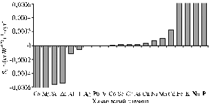
The toxic dose of cadmium introduced to a diet provoked a reliable elimination of Mg and Ca from the body (Fig.). Selenium content decreased by 52,8% (p ≤ 0,01), I - by 21,6% (p ≤ 0,05), Zn - by 12,3% (p ≤ 0,05), and Ag – by 50% (p £ 0,01). A corresponding S equaled to -0,0000140; -0,0000580; -0,0005500 and 0,0000001 g · (kg · W0,75)-1 · day-1. At the same time, there was observed a significant reduce in toxic elements pool: Al — by 41,3 % (р ≤ 0,01), Pb — by 33,4 % (р ≤ 0,01) and Sr — by 24,3 % (р ≤ 0,01) compared with control; S amounted to, respectively, -0,0001200; 0,0000017 and -0,0005600 g · (kg · W0,75)-1 · day-1.
Macroelements show the best plasticity under a toxic load in an organism. In this experiment, the diet with Cd contributed to the reduce in contents of Ca by 26,7%, K — by 22,6 % (р ≤ 0,05) and Mg — by 23,7 % vs. control while corresponding S amounted to -0,0248000; 0,0425900 and 0,0010000 g · (kg · W0,75)-1 · day-1.
Changes in elementary metabolism can be expressed by a ratio, where above and below the line are the elements whose content increased and decreased, respectively:
![]() .
.
 |
The rate (S) of accumulation (elimination) of chemical elements in rats fed the diets with toxic dose of cadmium. Denotations: |
Histological investigation of reproductive organs revealed in Cd-treated rats the inhibited ovulation and cystiform cavities developed instead of follicles.
In the second experiment, feed consumption in the control group for the accounted period averaged 29 554 g/bird, which was 0,4% higher than in the test group. However, the test group showed the increase in digestibility of organic matter by 4,8, fat – by 11,4, and carbohydrates – by 6,0% compared with control.
It is known that the deficit of chemical elements reduces the coefficient of performance (COP) of feed, disturbs animals’ reproductive function, health and productivity (9). The problem intensifies at a minimal income of macro- and microelements, which violates physiological functions of individual organs and a whole body.
Against the background of toxic doses of cadmium sulfate introduced with a diet, a total pool of toxic elements amounted to 0,0642000 mmol/g, which was 8,3% less than in the initial period (14 weeks). There was recorded a significant increase in contents of Al and Cd in tissues of reproductive organs of treated hens (34,1%) vs. control (24,7%) (p ≤ 0,05).
The content of conditionally essential elements in reproductive organs of the test group compared with 14-weeks age decreased from 0,26 to 0,18 mmol/g. They showed 53% higher content of arsenic (p ≤ 0,05) and 55,0% higher content of essential elements (2,02 mmol/g) relative to the control group.
In reproductive organs of Cd-treated hens the maximum contents were detected in respect of the essential elements iron and zinc (exceeding control by, resp., 63,1 and 45,0%; p ≤ 0,05) while the decrease in Cr and Se (resp., by 33,8 and 55,5%; p ≤ 0,05).
In the period of intense growth of reproductive organs under cadmium load, there was observed the increase in contents of K, Mg and P – respectively, by 46,4, 36,1 and 35,3% (p ≤ 0,05).
The ratio of Ca:P in the control group at the age of 14 was 1:14, but at the age of 16 weeks it reduced to 1:9. In the case of Cd-supplemented diet, it equaled to 1:26, which indirectly supports the fact of antagonistic interactions between cadmium and calcium.
A short-term feeding of high doses of cadmium was associated with significant differences in contents of chemical elements in treated hens compared with control:
![]() .
.
These changes show the violation of stromal-follicular autocrine and paracrine interactions along with a dysfunction in the basic control level, which can be associated with direct Cd-induced damage to hypothalamus and pituitary gland (10).
Thus, toxic doses of cadmium in the diet of laying hens and rats have resulted in disturbance of the animals’ elemental metabolism in a whole body as well as in its systems, which was accompanied by a decrease in contents of many essential elements (Mg, Ca, Se, I and Zn) and suppressed functional activity of reproductive organs.
REFERENCES
1. Eikhler V., Yady v nashei pische (Food and Poisons in Our Life), Moscow, pp. 54-55.
2. Topuria G.M. and Zhukov A.P., Contents of Heavy Metals in Agroecosystem Elements of East Orenburg, in Mat. Sibirskoi mezhd. nauch.-prakt. konf. “Aktual’nyevoprosyveterinarnoymeditsiny” (Proc. Siberian Int. Sci.-Pract. Congress “Topical Issues of Veterinary Medicine”), Ornburg, 2004, vol. 2, pp. 242-245.
3. Osintseva L.A., Motovilov K.Ya. and Korkina V.I., Accumulation of Heavy Metals in Bee-Farming Production, S.-kh. biol., 2010, no. 2, pp. 88-90.
4. Volkova N.A. and Karplyuk I.A., The Study of Mutagenic Action of Cadmium at Per Oral Introduction, Voprosy pitaniya, 1990, no. 3, pp. 74-76.
5. Es’kov E.K. and Kir’yakulov V.M., Contents of Heavy Metals in Tissues of Settled Ducks Wintering in Moscow Oblast’, S.-kh. biol., 2008, no. 6, pp. 115-118.
6. Hill C.H. and Hill F.W., Influence of High Levels of Minerals on the Susceptibility of Chicks to Salmonella gallinarum, J. Nutr., 1974, vol. 80, pp. 227-230.
7. Fisinin V.I., Imangulov Sh.A., Egorov I.A. et al., Normirovanie kormleniya sel’skokhozyaistvennoi ptitsy po dostupnym aminokislotam: metod. recom. (Norms of Available Amino Acids in Poultry Diets: Methodological Recommendations), Sergiev Posad, 2000.
8. Imangulov Sh.A., Egorov I.A., Okolelova T.M. et al., Metodika provedeniya nauchnykh i proizvodstvennykh issledovaniy po kormleniyu sel’skokhozyaistvennoi ptitsy: rekomendatsii (Recommendations for the Technique of Scientific and Applied Researches on Feeding Poultry), Sergiev Posad, 2004.
9. Anke M., Grun M. and Partschefeld M., The Essentiality of Arsenic for Animals, in TraceSubstances in Environmental Health, Hemphill D.D., Ed., Columbia, MO, 1976, vol. 10, pp. 403-409.
10. Miller R.H. and Bellinger D., Metalls, in Occuhat. Environ. Reprod. Hazards, Baltimore, 1993, pp. 233-252.
Orenburg State University, Orenburg 460018, Russia, |
Received May 12, 2010
|


